For UK patients exploring the fast-growing world of dental tourism, navigating international clinical environments can feel overwhelming. When traveling overseas to places like Hungary, Poland, or Croatia for significant procedures like full mouth reconstructions, advanced dental implants, or smile makeovers, a universal language binds your treatment together: maxillofacial radiology.
Many patients receive digital diagnostic reports filled with dense terminology like radiolucency, trabecular bone density, or alveolar resorption. Understanding these terms is vital. A patient guide titled “Understanding Your Dental X-rays and CT Scans: What Your Dentist Sees” serves as an educational bridge, helping patients move from a place of confusion to informed clarity.
As a dental tourism review expert, I have analyzed this guide’s content, clarity, and practical value. This comprehensive review examines how effectively the guide empowers UK patients to understand their diagnostic data, evaluate international treatment plans, and safely navigate the world of cross-border dentistry.
The Core Objective of the Guide
The primary goal of “Understanding Your Dental X-rays and CT Scans: What Your Dentist Sees” is to pull back the curtain on the black-and-white, highly technical imagery used in dental diagnostics. For dental tourists, this insight is a major asset.
When a patient seeks consultations from multiple clinics overseas, they often receive conflicting treatment plans. One clinic might recommend an immediate-load All-on-4 configuration, while another insists on bilateral sinus lifts and an All-on-6 framework. By breaking down exactly how dentists analyze bone volume, root health, and structural pathologies, this guide transforms patients into active, confident participants in their own care.
Key Technical Foundations Covered in the Guide
To help patients think like a clinician, the guide introduces the fundamental physics and biological interactions behind image formation in dental radiology.
The Concept of Radiodensity
The guide does an excellent job of simplifying how X-ray photons interact with the human body. It introduces patients to two foundational concepts that appear on every diagnostic report:
-
Radiopaque (White/Light Zones): High-density structures absorb the majority of the radiation beam, preventing it from reaching the digital sensor. The guide explains that healthy tooth enamel, dense cortical bone, metal crown frameworks, and root-filling materials (like gutta-percha) appear bright white on screen.
-
Radiolucent (Dark/Black Zones): Low-density structures or empty spaces allow X-ray beams to pass through easily, striking the digital receptor with high energy. Soft tissues, active dental infections (abscesses), the pulp chamber, nerve pathways, and areas of advanced bone loss all appear as dark shadows.
Understanding this simple contrast allows a UK patient to look at an image and easily spot a dark, shadow-like circle at the tip of a root, identifying it as a chronic periapical infection that must be resolved before placing an implant.
2D vs. 3D Dental Imaging Modalities
The guide provides a structured breakdown of the imaging modalities used throughout a typical dental tourism timeline, helping patients understand when and why each is necessary.
1. Intraoral Radiographs (Bitewings and Periapicals)
Intraoral imaging requires placing the digital sensor directly inside the patient’s mouth to capture highly localized, ultra-high-resolution views.
-
Bitewings: The guide explains that these are primarily used to identify interproximal dental decay (cavities hidden between teeth) and monitor early crestal bone loss caused by periodontal disease.
-
Periapicals: These capture the entire length of the tooth, from the visible crown down to the root tip and surrounding bone. The guide explains how dentists use these to evaluate root canal treatments or assess individual tooth stability before using them as anchor units for bridges.
2. Panoramic Radiographs (Orthopantomograms / OPG)
For dental tourists, the OPG is the standard entry requirement. It provides a broad, two-dimensional view of the entire upper and lower jaw, the maxillary sinuses, and the temporomandibular joints (TMJ) in a single image.
The guide highlights the convenience of OPGs for initial remote assessments, but it also notes their limitations, such as mild magnification and structural overlap. This clarifies why an overseas clinic can provide an initial quote based on an OPG, but will always require a 3D scan once the patient arrives at the clinic.
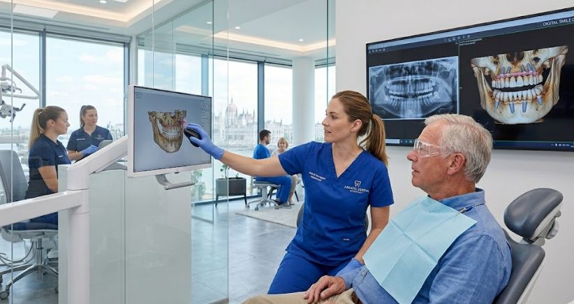
3. Cone Beam Computed Tomography (CBCT)
The guide dedicates a major section to Cone Beam Computed Tomography (CBCT), a technology that has redefined modern implant dentistry. Unlike conventional medical CT scans that use a fan-shaped beam and deliver higher radiation doses, a CBCT uses a cone-shaped beam to capture a full 3D model of the jaw in a single, fast rotation.
| Feature | Conventional Medical CT Scan | Cone Beam CT (CBCT) |
| X-ray Beam Geometry | Fan-shaped slice emission | Cone-shaped single volumetric sweep |
| Radiation Exposure | Significantly Higher | Markedly Lower |
| Primary Tissue Focus | Soft tissue and bone adaptation | High-resolution hard tissue structures |
| In-Clinic Availability | Requires dedicated hospital facilities | Compact design for dental offices |
The guide emphasizes that for full mouth restorations—such as All-on-4 or All-on-6 configurations—a 3D CBCT scan is essential. It allows surgeons to measure the exact height, width, and density of the available jawbone down to fractions of a millimeter. This precision ensures implants are placed safely without impacting vital structures like the inferior alveolar nerve or perforating the maxillary sinus cavity.
What the Dentist Sees: Evaluating Bone and Pathologies
The standout section of this guide explains how dentists analyze common oral pathologies and anatomical variations. For patients seeking dental care abroad, understanding this process helps them verify the medical necessity of additional procedures, like bone grafts or sinus lifts.
1. Alveolar Bone Resorption and Quality
When teeth are lost, the surrounding jawbone naturally begins to shrink due to a lack of chewing stimulation. The guide explains how a surgeon evaluates this on a CBCT scan.
If a patient has been missing their upper molars for years, the bone often resorbs vertically while the sinus cavity expands downward. The guide shows patients how this appears as a thin strip of white bone beneath a large dark space (the sinus). This visual explanation helps patients understand why a sinus lift is necessary to safely place an implant.
2. Identifying Active Periapical and Periodontal Disease
The guide helps patients identify signs of active infection on their scans:
-
Periapical Abscesses: These appear as dark, circular halos at the root tip, indicating that bacteria have destroyed the local bone matrix.
-
Periodontal Bone Loss: Instead of the bone sitting high around the neck of the tooth, the guide shows how chronic gum disease lowers the bone level, exposing the root forks. This explains why some teeth cannot be saved and must be extracted as part of a full mouth restoration.
3. Anatomical Boundaries: The Inferior Alveolar Nerve
In the lower jaw (mandible), the inferior alveolar nerve runs directly beneath the tooth roots. The guide details how dentists use 3D cross-sectional views to map this nerve canal. This mapping allows them to plan the precise length and tilt of the implants, ensuring they avoid any contact with the nerve that could cause permanent lip or chin numbness.
The Dental Tourism Perspective: Benefits for UK Patients
For UK patients traveling overseas for dental care, the insights provided by “Understanding Your Dental X-rays and CT Scans” offer significant advantages.
Avoid Over-Treatment and Ghost Clinics
The dental tourism market is highly competitive. While the majority of international clinics operate with high ethical and clinical standards, a few may suggest unnecessary extractions or excessive bone augmentations to increase treatment costs.
By understanding your own scans, you can ask targeted questions: “My CBCT shows 6mm of horizontal bone width at tooth 14; why is a block bone graft indicated rather than a conservative ridge split?” This level of detail shows international clinics that you are an informed patient, encouraging transparent communication.
Clear Communication Across Distance
When you return to the UK after the first stage of an implant procedure, your local dentist may handle your routine maintenance or check-ups. Having copies of your 3D CBCT scans and understanding the structural details allows you to coordinate care smoothly between your international surgeon and your local UK team, ensuring nothing is lost in translation.
Areas for Improvement within the Guide
While the guide is excellent, adding a few details could make it even more valuable for patients considering dental tourism:
-
A Guide to Radiation Dosages: Patients often worry about radiation safety when undergoing multiple OPG and CBCT scans during their treatment journey. The guide would benefit from contextualizing these numbers. For example, a modern digital dental CBCT scan typically exposes a patient to between $20\mu\text{Sv}$ and $100\mu\text{Sv}$ of radiation. This is minimal compared to a standard medical CT scan (which can exceed $2000\mu\text{Sv}$) or even the natural background radiation experienced during a round-trip flight from London to Budapest.
-
Addressing Artifacts from Metal Work: The guide should note that existing metal fillings, gold crowns, or older implants can create “scatter artifacts” or bright streaks on a 3D scan. Explaining this helps patients understand why a surgeon might need to take additional steps to get a clear view around older dental work.
Step-by-Step Diagnostic Timeline for Dental Tourists
To help patients apply this information practically, here is a breakdown of how imaging is typically utilized throughout an overseas full mouth restoration project:
Final Expert Verdict
“Understanding Your Dental X-rays and CT Scans: What Your Dentist Sees” is a highly valuable resource for any UK patient considering complex dental care abroad. It breaks down complex radiological physics into clear, practical insights, allowing patients to view their own diagnostic scans with confidence.
For dental tourists, this knowledge is a powerful asset. It allows you to verify your treatment needs, communicate clearly with international specialists, and ensure your full mouth restoration is built on a safe, well-planned foundation.
References
-
Alves, I. S., Vendramini, D. F. V., Leite, C. C., Gebrim, E. M. M. S., & Passos, U. L. (2021). Dental findings on face and neck imaging. Radiologia Brasileira, 54(2), 107-114. https://doi.org/10.1590/0100-3984.2019.0104
-
Alzaqy, H. N. (2024). Dental radiology in clinical practice: Principles, techniques, and diagnostic value. The Journal of Diabetic Studies, 21(1), 45-58.
-
Benavides, E. (2026). American Dental Association and American Academy of Oral and Maxillofacial Radiology patient selection for dental radiography and cone-beam computed tomography. Rhode Island Department of Health Medical Standards Guidelines, 1-14.
-
Code, S. (2022). Safety Code 30: Radiation Protection in Dentistry — Recommended Safety Procedures for the Use of Dental X-Ray Equipment. Environmental and Workplace Health Series, Canada.ca.
-
Scheinfeld, M. H., Shifteh, K., Avery, L. L., Dym, H., & Dym, R. J. (2012). Teeth: What radiologists should know. RadioGraphics, 32(7), 1927-1944. https://doi.org/10.1148/rg.327125717
-
Venkatesh, E., & Venkatesh Elluru, S. (2017). Cone beam computed tomography: Basics and applications in dentistry. Journal of Istanbul University Faculty of Dentistry, 51(3 Suppl 1), S102-S121. https://doi.org/10.17096/jiufd.00289
-
Yeung, A. W. K. (2023). Dental CT scan vs. Cone Beam CT: An overview. International Team for Implantology (ITI) Clinical Insights, Article 402.